
Knowledge check cardiovascular and respiratory disorders NURS 6501
Walden University Knowledge check cardiovascular and respiratory disorders NURS 6501-Step-By-Step Guide
This guide will demonstrate how to complete the Walden University Knowledge check cardiovascular and respiratory disorders NURS 6501 assignment based on general principles of academic writing. Here, we will show you the A, B, Cs of completing an academic paper, irrespective of the instructions. After guiding you through what to do, the guide will leave one or two sample essays at the end to highlight the various sections discussed below.
How to Research and Prepare for Knowledge check cardiovascular and respiratory disorders NURS 6501
Whether one passes or fails an academic assignment such as the Walden University Knowledge check cardiovascular and respiratory disorders NURS 6501 depends on the preparation done beforehand. The first thing to do once you receive an assignment is to quickly skim through the requirements. Once that is done, start going through the instructions one by one to clearly understand what the instructor wants. The most important thing here is to understand the required format—whether it is APA, MLA, Chicago, etc.
After understanding the requirements of the paper, the next phase is to gather relevant materials. The first place to start the research process is the weekly resources. Go through the resources provided in the instructions to determine which ones fit the assignment. After reviewing the provided resources, use the university library to search for additional resources. After gathering sufficient and necessary resources, you are now ready to start drafting your paper.
How to Write the Introduction for Knowledge check cardiovascular and respiratory disorders NURS 6501
The introduction for the Walden University Knowledge check cardiovascular and respiratory disorders NURS 6501 is where you tell the instructor what your paper will encompass. In three to four statements, highlight the important points that will form the basis of your paper. Here, you can include statistics to show the importance of the topic you will be discussing. At the end of the introduction, write a clear purpose statement outlining what exactly will be contained in the paper. This statement will start with “The purpose of this paper…” and then proceed to outline the various sections of the instructions.

Struggling to Meet Your Deadline?
Get your assignment on Knowledge check cardiovascular and respiratory disorders NURS 6501 done on time by medical experts. Don’t wait – ORDER NOW!
How to Write the Body for Knowledge check cardiovascular and respiratory disorders NURS 6501
After the introduction, move into the main part of the Knowledge check cardiovascular and respiratory disorders NURS 6501 assignment, which is the body. Given that the paper you will be writing is not experimental, the way you organize the headings and subheadings of your paper is critically important. In some cases, you might have to use more subheadings to properly organize the assignment. The organization will depend on the rubric provided. Carefully examine the rubric, as it will contain all the detailed requirements of the assignment. Sometimes, the rubric will have information that the normal instructions lack.
Another important factor to consider at this point is how to do citations. In-text citations are fundamental as they support the arguments and points you make in the paper. At this point, the resources gathered at the beginning will come in handy. Integrating the ideas of the authors with your own will ensure that you produce a comprehensive paper. Also, follow the given citation format. In most cases, APA 7 is the preferred format for nursing assignments.
How to Write the Conclusion for Knowledge check cardiovascular and respiratory disorders NURS 6501
After completing the main sections, write the conclusion of your paper. The conclusion is a summary of the main points you made in your paper. However, you need to rewrite the points and not simply copy and paste them. By restating the points from each subheading, you will provide a nuanced overview of the assignment to the reader.
How to Format the References List for Knowledge check cardiovascular and respiratory disorders NURS 6501
The very last part of your paper involves listing the sources used in your paper. These sources should be listed in alphabetical order and double-spaced. Additionally, use a hanging indent for each source that appears in this list. Lastly, only the sources cited within the body of the paper should appear here.
Stuck? Let Us Help You
Completing assignments can sometimes be overwhelming, especially with the multitude of academic and personal responsibilities you may have. If you find yourself stuck or unsure at any point in the process, don’t hesitate to reach out for professional assistance. Our assignment writing services are designed to help you achieve your academic goals with ease.
Our team of experienced writers is well-versed in academic writing and familiar with the specific requirements of the Knowledge check cardiovascular and respiratory disorders NURS 6501 assignment. We can provide you with personalized support, ensuring your assignment is well-researched, properly formatted, and thoroughly edited. Get a feel of the quality we guarantee – ORDER NOW.
Sample Answer for Knowledge check cardiovascular and respiratory disorders NURS 6501 Included After Question
Knowledge check cardiovascular and respiratory disorders NURS 6501 Knowledge check cardiovascular and respiratory disorders NURS 6501
Scenario 5: COPD
A 66-year-old female with a 50 pack/year history of cigarette smoking had a CT scan and was diagnosed with emphysema. He asks if this means he has chronic obstructive pulmonary disease (COPD).
Question:
1. There is a clear relationship between emphysema and COPD, explain the pathophysiology of emphysema and the relationship to COPD.
Your Answer:
Emphysema is a condition affecting the airways, characterized by the permanent enlargement of the air exchange pathways. It is associated with the destruction of alveolar walls, which do not exhibit fibrosis. Chronic exposure to irritants triggers the recruitment of neutrophils, macrophages, and lymphocytes to the lungs, leading to progressive damage caused by inflammatory oxidative stress. In emphysema, the destruction of alveoli results in a reduced surface area available for efficient gas exchange. Consequently, this leads to a significant mismatch between ventilation and perfusion, impacting the overall respiratory function.
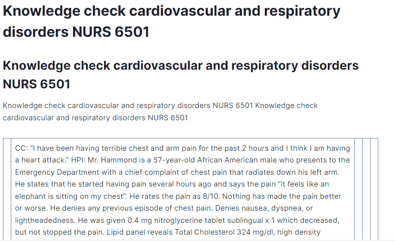
| CC: “I have been having terrible chest and arm pain for the past 2 hours and I think I am having a heart attack.” HPI: Mr. Hammond is a 57-year-old African American male who presents to the Emergency Department with a chief complaint of chest pain that radiates down his left arm. He states that he started having pain several hours ago and says the pain “it feels like an elephant is sitting on my chest”. He rates the pain as 8/10. Nothing has made the pain better or worse. He denies any previous episode of chest pain. Denies nausea, dyspnea, or lightheadedness. He was given 0.4 mg nitroglycerine tablet sublingual x 1 which decreased, but not stopped the pain. Lipid panel reveals Total Cholesterol 324 mg/dl, high density lipoprotein (HDL) 31 mg/dl, Low Density Lipoprotein (LDL) 122 mg/dl, Triglycerides 402 mg/dl, Very Low-Density Lipoprotein (VLDL) 54 mg/dl His diagnosis is an acute inferior wall myocardial infarction. 1 of 2 Questions: Why is HDL considered the “good” cholesterol? | ||||
| Selected Answer: HDL carries between 20-25% of the total cholesterol in plasma. It is a good cholesterol since it accumulates the excess cholesterol that exists in body cells for excretion in the liver. Correct Answer: HDL is considered the good cholesterol because it collects excess cholesterol in the body cells and transports it to the liver where it is excreted in the body cells and transports it to the liver where it is excreted in the body. HDL carries 20-25% of total plasma cholesterol. Response Feedback: [None Given] |
Question 2
3 out of 3 points
| CC: “I have been having terrible chest and arm pain for the past 2 hours and I think I am having a heart attack.” HPI: Mr. Hammond is a 57-year-old African American male who presents to the Emergency Department with a chief complaint of chest pain that radiates down his left arm. He states that he started having pain several hours ago and says the pain “it feels like an elephant is sitting on my chest”. He rates the pain as 8/10. Nothing has made the pain better or worse. He denies any previous episode of chest pain. Denies nausea, dyspnea, or lightheadedness. He was given 0.4 mg nitroglycerine tablet sublingual x 1 which decreased, but not stopped the pain. Lipid panel reveals Total Cholesterol 324 mg/dl, high density lipoprotein (HDL) 31 mg/dl, Low Density Lipoprotein (LDL) 122 mg/dl, Triglycerides 402 mg/dl, Very Low-Density Lipoprotein (VLDL) 54 mg/dl His diagnosis is an acute inferior wall myocardial infarction. 2 of 2 Questions: Explain the role inflammation has in the development of atherosclerosis. | ||||
| Selected Answer: Mitochondrial damage caused by chronic inflammatory processes cause inflammation in the heart muscles. These processes cause an increase in the production of free radicals that activate the continuous cycle of chronic inflammation. Correct Answer: Inflammation in the heart muscle caused by chronic inflammatory processes leads to mitochondrial damage that results in an increased free radical production that further activates the chronic inflammatory vicious cycle. Response Feedback: [None Given] | ||||
Cardiovascular and Respiratory Disorders
In this exercise, you will complete a 5-essay type question Knowledge Check to gauge your understanding of this module’s content.
Possible topics covered in this Knowledge Check include:
- myocardial infarction
- endocarditis
- myocarditis
- valvular disorders
- lipid panels
- coagulation
- clotting cascade
- deep vein thrombosis
- hypertension
- heart failure
- COPD
- asthma
- pneumonias
Resources
Be sure to review the Learning Resources before completing this activity.
Click the weekly resources link to access the resources.
WEEKLY RESOURCES
By Day 7 of Week 3
Complete the Knowledge Check by Day 7.
This quiz was locked Mar 19 at 10:59pm.
Attempt History
| Attempt | Time | Score | |
| LATEST | Attempt 1 | 1,270 minutes | 20 out of 20 |
Score for this quiz: 20 out of 20
Submitted Mar 16 at 8:41am
This attempt took 1,270 minutes.
Question 1
4 / 4 pts
Scenario 1: Myocardial Infarction
CC: “I woke up this morning at 6 a.m. with numbness in my left arm and pain in my chest. It feels tight right here (mid-sternal).” “My dad had a heart attack when he was 56-years-old and I am scared because I am 56-years-old.”
HPI: Patient is a 56-year-old Caucasian male who presents to Express Hospital Emergency Department with a chief complaint of chest pain that radiates down his left arm. He states this started this morning and has been getting worse, pointing to the mid-sternal area, “it feels like an elephant is sitting on my chest and having a hard time breathing”. He rates the pain as 9/10. Nothing has made the pain better or worse. He denies any previous episode of chest pain. Denies nausea, or lightheadedness. Nitroglycerin 0.4 mg tablet sublingual x 1 which decreased pain to 7/10.
Lipid panel reveals Total Cholesterol 424 mg/dl, high density lipoprotein (HDL) 26 mg/dl, Low Density Lipoprotein (LDL) 166 mg/dl, Triglycerides 702 mg/dl, Very Low-Density Lipoprotein (VLDL) 64 mg/dl
His diagnosis is an acute inferior wall myocardial infarction.
A Sample Answer For the Assignment: Knowledge check cardiovascular and respiratory disorders NURS 6501
Title: Knowledge check cardiovascular and respiratory disorders NURS 6501
Question:
Which cholesterol is considered the “good” cholesterol and what does it do?
Your Answer:
This case study is about a 56-year-old patient that presents to Express Hospital Emergency Department with a chief complaint of chest pain radiating to his left arm. The pain started this morning and has been worsening, pointing to the mid-sternal area. The chest pain is rated at 9/10 and nitroglycerin tablet has decreased it to 7/10. A diagnosis of acute wall myocardial infarction has been made.
Cholesterols exist in different types. They include low-density lipoproteins and high-density lipoproteins. Low density lipoproteins are the bad proteins while high density lipoproteins are the good proteins. High density lipoproteins are the good proteins because they absorb cholesterol from the contained in the blood and carries them to the liver for elimination. The benefit of high density lipoproteins is that it lowers the risk of an individual developing cardiovascular problems such as stroke, hypertension, arteriosclerosis, and heart failure (CDC, 2022).
Low triglyceride levels in combination with high density lipoproteins are beneficial in lowering the risk of cardiovascular problems. High density lipoproteins also prevent the development of plaques in the arteries. This lowers the threshold of arteriosclerotic plaques in healthy individuals. High density lipoproteins are also not associated with inflammation due to oxidative processes (Brandts & Ray, 2020). As a result, they protect and preserve the health and survival or cells and artery walls.
In summary, high-density lipoproteins are the good proteins while low density lipoproteins are the bad ones. High density lipoproteins lower the individual risk of developing cardiovascular complications. Together with low triglyceride levels, high density lipoproteins protect the integrity of arterial walls by preventing development of plaques and oxidative processes that cause inflammation.
References
Brandts, J., & Ray, K. K. (2020). Low Density Lipoprotein Cholesterol–Lowering Strategies and Population Health. Circulation, 141(11), 873–876. https://doi.org/10.1161/CIRCULATIONAHA.119.043406
- (2022, October 24). LDL and HDL Cholesterol and Triglycerides | cdc.gov. Centers for Disease Control and Prevention. https://www.cdc.gov/cholesterol/ldl_hdl.htm
Question 2
4 / 4 pts
Scenario 1: Myocardial Infarction
CC: “I woke up this morning at 6 a.m. with numbness in my left arm and pain in my chest. It feels tight right here (mid-sternal).” “My dad had a heart attack when he was 56-years-old and I am scared because I am 56-years-old.”
HPI: Patient is a 56-year-old Caucasian male who presents to Express Hospital Emergency Department with a chief complaint of chest pain that radiates down his left arm. He states this started this morning and has been getting worse, pointing to the mid-sternal area, “it feels like an elephant is sitting on my chest and having a hard time breathing”. He rates the pain as 9/10. Nothing has made the pain better or worse. He denies any previous episode of chest pain. Denies nausea, or lightheadedness. Nitroglycerin 0.4 mg tablet sublingual x 1 which decreased pain to 7/10.
Lipid panel reveals Total Cholesterol 424 mg/dl, high density lipoprotein (HDL) 26 mg/dl, Low Density Lipoprotein (LDL) 166 mg/dl, Triglycerides 702 mg/dl, Very Low-Density Lipoprotein (VLDL) 64 mg/dl
His diagnosis is an acute inferior wall myocardial infarction.
Question:
1. How does inflammation contribute to the development of atherosclerosis?
Your Answer:
This scenario is a continuation from scenario 1. The patient has been diagnosed with acute inferior wall myocardial infection. This is after he presented with complaints that included chest pain radiating to his left arm worsening in nature and pain responsive to nitroglycerin.
Inflammatory processes contribute to the development of atherosclerosis. The first step in the process is the deposition of low density lipoproteins in the walls of the arteries. Elevated levels of triglycerides accelerate the deposition of low-density lipoproteins in the intima of the arteries. The deposition initiates a series of cascades that contribute to the development of atherosclerosis. First, there is the activation of the arteriole endothelium with the deposition. This promotes the sticking or adhesion of molecules in the walls of the arteries. There is also the recruitment of T cells and monocytes under the influence of chemokines. The released monocytes differentiate into macrophages, which are involved in the upregulation of pattern recognition receptors such as the scavenger receptors. The next step is the lipoprotein internalization under the influence of scavenger receptors, which lead to the formation of foam cells. Toll-like receptors stimulate the release of inflammatory cells such as proteases, vasoactive molecules and cytokines. These cells stimulate inflammation in the walls of the arteries and growth of the atherosclerotic plaques (Libby, 2021; Malekmohammad et al., 2021; Soehnlein & Libby, 2021). In severe inflammation, plaque rupture and proteolysis may occur leading to infarction, ischemia, and thrombus formation.
In summary, inflammatory processes contribute to atherosclerosis. The process begins with the deposition of low-density lipoproteins on the walls of the arteries. The deposition initiates a series of processes that lead to inflammation. Severe inflammation can cause plaque rupture, which leads to infarction, thrombus formation, and ischemia.
References
Libby, P. (2021). Inflammation in Atherosclerosis—No Longer a Theory. Clinical Chemistry, 67(1), 131–142. https://doi.org/10.1093/clinchem/hvaa275
Malekmohammad, K., Bezsonov, E. E., & Rafieian-Kopaei, M. (2021). Role of Lipid Accumulation and Inflammation in Atherosclerosis: Focus on Molecular and Cellular Mechanisms. Frontiers in Cardiovascular Medicine, 8. https://www.frontiersin.org/articles/10.3389/fcvm.2021.707529
Soehnlein, O., & Libby, P. (2021). Targeting inflammation in atherosclerosis—From experimental insights to the clinic. Nature Reviews Drug Discovery, 20(8), Article 8. https://doi.org/10.1038/s41573-021-00198-1
Question 3
4 / 4 pts
Scenario 2: Pleural Friction Rub
A 35-year-old female with a positive history of systemic lupus erythematosus (SLE) presents to the Emergency Room (ER) with complaints of sharp retrosternal chest pain that worsens with deep breathing or lying down. She reports a 5-day history of low-grade fever, listlessness and says she feels like she had the flu. Physical exam reveals tachycardia and a pleural friction rub. She was diagnosed with acute pericarditis.
Question:
1. Because of the result of a pleural friction rub, what does the APRN recognize?
Your Answer:
This scenario is about a 35-year-old female patient that has been diagnosed with acute pericarditis. The patient has a positive history of systemic lupus erythematosus (SLE). She presents to the emergency room with complaints of sharp retrosternal chest pain that worsens with deep breathing or lying down. She reports a history low grade fever for the last five days.
The advanced practice registered nurser recognizes that a pleural friction rub is among the adventitious sounds that indicate an underlying pathology. The rub results from inflammation and roughened pleural surfaces that rub against each other during breathing. The nurse would suspect causes such as serositis, pleuritis, or pleural effusion. The nurse would also infer that there is a reduced production of pleural fluid by the pleura, increasing the friction between pleural membranes. The accompanying symptoms would include pleuritic chest pain, which may be referred to the shoulder or the neck if it is near the diaphragm. Pain sensation the client reports is due to the somatic nerves that innervate the parietal pleural (Adderley & Sharma, 2022; Moriki et al., 2023). The nurse should anticipate prescribing analgesics and treating the cause.
In summary, the pleural friction rub is attributed to inflammatory process and reduced production of pleural fluid, causing friction. Patients experience sharp sternal pain during breathing or when lying down. The appropriate treatment will aim at relieving symptoms and treating the causative agent.
References
Adderley, N., & Sharma, S. (2022). Pleural Friction Rub. In StatPearls. StatPearls Publishing. http://www.ncbi.nlm.nih.gov/books/NBK537118/
Moriki, D., Koumpagioti, D., Kalogiannis, M., Sardeli, O., Galani, A., Priftis, K. N., & Douros, K. (2023). Physicians’ ability to recognize adventitious lung sounds. Pediatric Pulmonology, 58(3), 866–870. https://doi.org/10.1002/ppul.26266
Question 4
4 / 4 pts
Scenario 4: Deep Venous Thrombosis (DVT)
A 81-year-old obese female patient who 48 hours post-op left total hip replacement. The patient has had severe nausea and vomiting and has been unable to go to physical therapy. Her mucus membranes are dry. The patient says the skin on her left leg is too tight. Exam reveals a swollen, tense, and red colored calf. The patient has a duplex ultrasound which reveals the presence of a deep venous thrombosis (DVT).
Question:
1. Given the history of the patient explain what contributed to the development of a deep venous thrombosis (DVT)
Your Answer:
This scenario is about an 81-year-old female patient that has been diagnosed with deep vein thrombosis. The patient is obese. She underwent left total hip replacement surgery 48 hours ago. She had severe nausea and vomiting, which has made it difficult for her to go for physical therapy. Her mucus membranes are dry. She report that her left leg is too tight, with physical examination revealing a swollen, red colored, and tense calf. Therefore, this essay examines the factors that contributed to the development of DVT.
Several factors are attributed to the development of DVT in the patient. One of them is immobility. Immobility increases the risk of DVT due to blood stasis. The patient in the case study has been bedridden because of severe nausea and vomiting. The other risk factor is surgery. Trauma in forms such as surgery increases the risk of blood clot formation within the blood vessels, leading to an increased risk of DVT. Surgeries of the legs and upper extremities have the highest risk of DVT. The patient is 48 hours post left total hip replacement surgery, hence, the risk.
The other risk factor that increased the risk of patient developing DVT is dehydration. Dehydration increases blood viscosity. The implication is the sluggish blood flow from the extremities, which increase the risk of DVT. The patient in the case study has experienced severe nausea and vomiting. Her mucus membranes are also dry, which affects blood viscosity. The other risk factor is obesity. Obesity increases the risk of inflammation within the blood vessels due to the formation of plaques. The inflammation increases the risk of blood clotting (Wang et al., 2019; Yu et al., 2020; Zhang et al., 2019). Obesity is also a risk factor for other comorbidities such s diabetes, which are associated with the increased risk of DVT.
In summary, the patient in the case study has several risk factors for DVT. They include her being obese, surgery, immobility, and dehydration. Therefore, prophylactic interventions should be adopted to prevent complications.
References
Wang, P., Kandemir, U., Zhang, B., Wang, B., Li, J., Zhuang, Y., Wang, H., Zhang, H., Liu, P., & Zhang, K. (2019). Incidence and Risk Factors of Deep Vein Thrombosis in Patients With Pelvic and Acetabular Fractures. Clinical and Applied Thrombosis/Hemostasis, 25, 1076029619845066. https://doi.org/10.1177/1076029619845066
Yu, Y., Tu, J., Lei, B., Shu, H., Zou, X., Li, R., Huang, C., Qu, Y., & Shang, Y. (2020). Incidence and Risk Factors of Deep Vein Thrombosis in Hospitalized COVID-19 Patients. Clinical and Applied Thrombosis/Hemostasis, 26, 1076029620953217. https://doi.org/10.1177/1076029620953217
Zhang, W., Huai, Y., Wang, W., Xue, K., Chen, L., Chen, C., & Qian, A. (2019). A Retrospective cohort study on the risk factors of deep vein thrombosis (DVT) for patients with traumatic fracture at Honghui Hospital. BMJ Open, 9(3), e024247. https://doi.org/10.1136/bmjopen-2018-02424
Question 5
4 / 4 pts
Scenario 5: COPD
A 66-year-old female with a 50 pack/year history of cigarette smoking had a CT scan and was diagnosed with emphysema. He asks if this means he has chronic obstructive pulmonary disease (COPD).
Question:
1. There is a clear relationship between emphysema and COPD, explain the pathophysiology of emphysema and the relationship to COPD.
Your Answer:
Emphysema is a respiratory condition characterized by the reduction in pulmonary elasticity recoil. The reduction results in air trapping within the lungs and increase in the lung volume beyond the normal level. These changes alter the physiological process of air exchange in the pleura and alveoli, leading to the compression of the airways. In addition, there is the reduction in airflow during the tidal and forced expiration. Consequently, there is the destruction in the matching of the required ventilation by the body with the alveolar pressure, leading to respiratory collapse. A relationship between emphysema and COPD exists. Emphysema is a type of COPD. However, patients may be diagnosed with COPD and not have emphysema (Amariei et al., 2019; Leap et al., 2021). Emphysema precipitates symptoms of COPD due to the damage to the alveoli, which makes them lose elasticity, hence, air trapping.
In summary, emphysema develops from the damage to the alveoli. The damage results in the loss of lung elasticity. Emphysema is related to COPD. The mechanisms in emphysema produce symptoms related to those of COPD.
References
Amariei, D. E., Dodia, N., Deepak, J., Hines, S. E., Galvin, J. R., Atamas, S. P., & Todd, N. W. (2019). Combined Pulmonary Fibrosis and Emphysema: Pulmonary Function Testing and a Pathophysiology Perspective. Medicina, 55(9), Article 9. https://doi.org/10.3390/medicina55090580
Leap, J., Arshad, O., Cheema, T., & Balaan, M. (2021). Pathophysiology of COPD. Critical Care Nursing Quarterly, 44(1), 2. https://doi.org/10.1097/CNQ.0000000000000334
Review Test Submission: Module 2 Knowledge Check
2 out of 2 points
| CC: “I have been having terrible chest and arm pain for the past 2 hours and I think I am having a heart attack.” HPI: Mr. Hammond is a 57-year-old African American male who presents to the Emergency Department with a chief complaint of chest pain that radiates down his left arm. He states that he started having pain several hours ago and says the pain “it feels like an elephant is sitting on my chest”. He rates the pain as 8/10. Nothing has made the pain better or worse. He denies any previous episode of chest pain. Denies nausea, dyspnea, or lightheadedness. He was given 0.4 mg nitroglycerine tablet sublingual x 1 which decreased, but not stopped the pain. Lipid panel reveals Total Cholesterol 324 mg/dl, high density lipoprotein (HDL) 31 mg/dl, Low Density Lipoprotein (LDL) 122 mg/dl, Triglycerides 402 mg/dl, Very Low-Density Lipoprotein (VLDL) 54 mg/dl His diagnosis is an acute inferior wall myocardial infarction. 1 of 2 Questions: Why is HDL considered the “good” cholesterol? | ||||
| Selected Answer: HDL carries between 20-25% of the total cholesterol in plasma. It is a good cholesterol since it accumulates the excess cholesterol that exists in body cells for excretion in the liver. Correct Answer: HDL is considered the good cholesterol because it collects excess cholesterol in the body cells and transports it to the liver where it is excreted in the body cells and transports it to the liver where it is excreted in the body. HDL carries 20-25% of total plasma cholesterol. Response Feedback: [None Given] | ||||
Question 2
3 out of 3 points
| CC: “I have been having terrible chest and arm pain for the past 2 hours and I think I am having a heart attack.” HPI: Mr. Hammond is a 57-year-old African American male who presents to the Emergency Department with a chief complaint of chest pain that radiates down his left arm. He states that he started having pain several hours ago and says the pain “it feels like an elephant is sitting on my chest”. He rates the pain as 8/10. Nothing has made the pain better or worse. He denies any previous episode of chest pain. Denies nausea, dyspnea, or lightheadedness. He was given 0.4 mg nitroglycerine tablet sublingual x 1 which decreased, but not stopped the pain. Lipid panel reveals Total Cholesterol 324 mg/dl, high density lipoprotein (HDL) 31 mg/dl, Low Density Lipoprotein (LDL) 122 mg/dl, Triglycerides 402 mg/dl, Very Low-Density Lipoprotein (VLDL) 54 mg/dl His diagnosis is an acute inferior wall myocardial infarction. 2 of 2 Questions: Explain the role inflammation has in the development of atherosclerosis. | ||||
| Selected Answer: Mitochondrial damage caused by chronic inflammatory processes cause inflammation in the heart muscles. These processes cause an increase in the production of free radicals that activate the continuous cycle of chronic inflammation. Correct Answer: Inflammation in the heart muscle caused by chronic inflammatory processes leads to mitochondrial damage that results in an increased free radical production that further activates the chronic inflammatory vicious cycle. Response Feedback: [None Given] | ||||
Question 3
1 out of 1 points
| A 45-year-old woman with a history of systemic lupus erythematosus (SLE) presents to the Emergency Room (ER) with complaints of sharp retrosternal chest pain that worsens with deep breathing or lying down. She reports a 3-day history of low-grade fever, listlessness and says she feels like she had the flu. Physical exam reveals tachycardia and a pleural friction rub. She was diagnosed with acute pericarditis. Question: What does the Advanced Practice Registered Nurse (APRN) recognize as the result of the pleural friction rub? | ||||
| Selected Answer: The pericardium tends to roughen following inflammation caused by a post-viral syndrome or an underlying autoimmune disease. The roughening is what produces a classic rub that an APRN can hear at the left sternal border and apex of the heart. Correct Answer: The inflammation of the pericardium, due to either the underlying autoimmune disease or a post viral syndrome, causes roughening of the pericardium. The roughening of the pericardium causes the classic “rub” which can best be heard at the apex of the heart and left sternal border. Response Feedback: [None Given] | ||||
Question 4
1 out of 1 points
| A 15-year-old adolescent male comes to the clinic with his parents with a chief complaint of fever, nausea, vomiting, poorly localized abdominal pain, arthralgias, and “swollen lymph nodes”. States he has felt “lousy” for a couple weeks. The fevers have been as high as 102 F. His parents thought he had the flu and took him to an Urgent Care Center. He was given Tamiflu® and sent home. He says the Tamiflu didn’t seem to work. States had a slight sore throat a couple weeks ago and attributed it to the flu. Physical exam revealed thin young man who appears to be uncomfortable but not acutely ill. Posterior pharynx reddened and tonsils 3+ without exudate. + anterior and posterior cervical lymphadenopathy. Tachycardic and a new onset 2/6 high-pitched, crescendo-decrescendo systolic ejection murmur auscultated at the left sternal border. Rapid strep +. The patient was diagnosed with acute rheumatic heart disease (RHD). Question: Explain how a positive strep test has caused the patient’s symptoms. | ||||
| Selected Answer: A pharyngeal infection with GABHs (Group A Beta Hemolytic Streptococcus) often leads to the development of RHD, which is an abnormal response to cell-mediated responses. The inflammatory cascade associated with this process cause exudative and proliferative lesions, and scarring in the valve tissue. Since it is the endocardium which contains valves that is primarily affected, inflammation of the endocardium results to subsequent inflammation of valves. Correct Answer: Rheumatic Heart Disease (RHD) only develops after a pharyngeal infection with Group A beta hemolytic streptococcus. It is an abnormal response to humoral and cell-mediated response to M proteins on the microorganisms. The intense inflammation caused by these reactions cause proliferative and exudative lesions in connective tissue. This inflammation causes scarring of the valve tissue. The inflammation usually affects the endocardium which contains the valves. Endocardial inflammation causes swelling of leaflets in the valves. Response Feedback: [None Given] | ||||
Question 5
1 out of 1 points
| The APRN sees a 74-year-old obese female patient who is 2 days post-op after undergoing left total hip replacement. The patient has had severe post op nausea and vomiting and has been unable to go to physical therapy. Her mucus membranes are dry. The patient says she feels like the skin on her left leg is too tight. Exam reveals a swollen, tense, and red colored calf. The patient has a duplex ultrasound which reveals the presence of a deep venous thrombosis (DVT). Question: Describe the factors that could have contributed to the development of a DVT in this patient explain how each of the factors could cause DVT. | ||||
| Selected Answer: When there is injury to a blood vessel, there is platelet adherence to blood vessel walls influenced by antiplatelet substances. When platelets aggregate, they form clots. Therefore, virchow’s triad damaged the blood vessel walls. Since this patient is reportedly obese, inability to engage in physical therapy and an advanced age caused venous stasis, Correct Answer: Virchow’s Triad caused damage to the walls of the vessels. When there is injury to the intimal layer of the vessel, antiplatelet substances such as nitric oxide and prostacyclin, along with the expression of collagen on the vessel wall, causes adherence of the platelets to the vessel wall. The platelets become activated then aggregate forming clots. Venous stasis as a result of obesity, patient’s advanced age and inability to go to physical therapy. Response Feedback: [None Given] | ||||
Question 6
1 out of 1 points
| A 45-year-old woman is 10 days status post partial small bowel resection for Crohn Disease and has been recuperating at home. She suddenly develops severe shortness of breath, becomes weak, and her blood pressure drops to 80/40 mmHg (previous readings ~130/80s mmHg). The pulse oximetry is 89% on room air. The APRN suspects the patient experienced a massive pulmonary embolus. Question: Explain why a large pulmonary embolus interferes with oxygenation. | ||||
| Selected Answer: The pulmonary embolus lodged itself strategically in pulmonary circulation resulting to a mismatch in ventilation/perfusion (V/Q) reducing the area for the exchange of oxygen. Correct Answer: The embolus lodges somewhere in the pulmonary circulation and causes a ventilation/perfusion mismatch (V/Q). Ventilation Perfusion mismatch or “V/Q defects” are defects in total lung ventilation perfusion ratio. It is a condition in which one or more areas of the lung receive oxygen but no blood flow, or they receive blood flow but no oxygen due to obstruction somewhere in the pulmonary circulation. This causes a decreased area for oxygen exchange. Response Feedback: [None Given] | ||||
Question 7
1 out of 1 points
| A 45-year-old woman is 10 days status post partial small bowel resection for Crohn Disease and has been recuperating at home. She suddenly develops severe shortness of breath, becomes weak, and her blood pressure drops to 80/40 mmHg (previous readings ~130/80s mmHg). The pulse oximetry is 89% on room air. While waiting for the Emergency Medical Service (EMS) to arrive, the APRN places EKG leads and the EKG demonstrates right ventricular strain. Question: Explain why a large pulmonary embolism causes right ventricular strain. | ||||
| Selected Answer: The ventilation/perfusion mismatch caused the release of inflammatory mediators resulting to vasoconstriction of the pulmonary system obstructing oxygenation and subsequent hypertension. This causes atelectasis and makes pumping of blood by the right ventricle difficult Correct Answer: The V/Q mismatch causes release of neurohumeral substances and inflammatory mediators that cause vasoconstriction of the pulmonary vasculature further impeding oxygenation. Hemodynamically, this vasoconstriction results in pulmonary hypertension, making it difficult for the right ventricle to pump blood. The V/Q mismatch also causes decreased production of surfactant causing atelectasis that further decreases surface area available for oxygen exchange. Response Feedback: [None Given] | ||||
Question 8
2 out of 2 points
| A 12-year-old girl is brought to the Emergency Room (ER) by her mother with complaints of shortness of breath, wheezing, tachypnea, tachycardia, and a non-productive cough. The mother states they had just come from a fall festival where the entire family enjoyed a hayride. The symptoms began shortly after they left the festival but got better a couple hours after they returned home. The symptoms began again about 6 hours later and seem to be worse. The mother states there is no history of allergies or frequent respiratory infections. The child is up to date on all vaccinations. The child was diagnosed with asthma. The nurse practitioner explained to the mother that her child was exhibiting symptoms of asthma, and probably had an early asthmatic response and a late asthmatic response. Question 1 of 2: Explain early asthmatic responses and the cells responsible for the responses. | ||||
| Selected Answer: There is an adaptive and innate immune response following an initial airway exposure to an antigen. Basophils, T helper cells, eosinophils, dendritic cells, and mast cells can initiate the inflammatory process. This process can peak at thirty minutes and resolve after three hours. Correct Answer: When there is an initial airway exposure to an antigen, an innate and adaptive immune response is initiated. Cells that can initiate the inflammation of the bronchial mucosa and hyperresonance of the airways include dendritic cells, T helper 2 lymphocytes, B lymphocytes, mast cells, neutrophils, eosinophils, and basophils. Early asthmatic response is a phase of bronchospasm that peaks at about 30 minutes and usually resolves after about 3 hours. Response Feedback: [None Given] | ||||
Question 9
1.8 out of 2 points
| A 12-year-old girl is brought to the Emergency Room (ER) by her mother with complaints of shortness of breath, wheezing, tachypnea, tachycardia, and a non-productive cough. The mother states they had just come from a fall festival where the entire family enjoyed a hayride. The symptoms began shortly after they left the festival but got better a couple hours after they returned home. The symptoms began again about 6 hours later and seem to be worse. The mother states there is no history of allergies or frequent respiratory infections. The child is up to date on all vaccinations. The child was diagnosed with asthma. The nurse practitioner explained to the mother that her child was exhibiting symptoms of asthma, and probably had an early asthmatic response and a late asthmatic response. Question 2 of 2: Explain late asthmatic responses and the cells responsible for the responses. | ||||
| Selected Answer: Early exposure in the initial phase mediate late asthma responses resulting to the release of inflammatory mediators such as prostaglandins D and leukotrienes with subsequent edema, bronchospasms, and secretion of mucus that obstruct the flow of air. With continuous obstruction, resistance sets in and air is trapped hence reduced lung perfusion and ventilation. Correct Answer: Late asthmatic responses are mediated by earlier exposure in early phase that causes a latent release of inflammatory mediators. These mediators, leukotrienes and prostaglandin D, cause bronchospasm, edema, and mucus secretions that obstruct airflow. Airway obstruction creates resistance to airflow and causes air trapping. Continued air trapping increases intrapleural and alveolar gas pressure, decreases ventilation and perfusion leading to uneven and variable ventilation/perfusion in the lung Response Feedback: timing of phase | ||||
Question 10
2 out of 2 points
| A 64-year-old man with a 40 pack/year history of cigarette smoking has been diagnosed with emphysema. He asks the APRN if this means he has COPD. Question 1 of 2: Explain the pathophysiology of emphysema and how it relates to COPD. | ||||
| Selected Answer: Emphysema causes the permanent airway enlargement characterized by damage to alveolar walls. With continuous exposure to irritants, there is an inflammatory oxidative stress involving lymphocytes, neutrophils, and macrophages causing more alveoli damage. This process reduces the surface area required for the exchange of gases with a significant ventilation/perfusion mismatch. Correct Answer: Emphysema is a disease of the airways that causes permanent enlargement of the gasexchange airways. It is accompanied by destruction of the alveolar walls do not appear to be fibrotic. Chronic exposure to irritants recruit neutrophils, macrophages, and lymphocytes to the lung resulting in progressive damage from inflammatory oxidative stress. Emphysema is characterized by destruction of alveoli leading to decreased surface area for gas exchange that causes significant ventilation/perfusion mismatch. Response Feedback: [None Given] | ||||
Question 11
2 out of 2 points
| A 64-year-old man with a 40 pack/year history of cigarette smoking has been diagnosed with emphysema. He asks the APRN if this means he has COPD. Question 2 of 2: Explain the pathophysiology of chronic bronchitis and how it relates to COPD. | ||||
| Selected Answer: The bronchi becomes inflamed when an individual inhales irritants. Inflammation increases the number and size of goblet cells and mucus glands, causes edema of the bronchial, and hypertrophy of the smooth muscles. Over time, the airway undergoes fibrosis and narrows. Since the functioning of the ciliary is also impaired, continuous mucus production hinders the patient’s ability to cough. In the advanced stages, both the large and small airways get involved, with an obstructed airflow during expiration that can result to a VQ mismatch. Correct Answer: Chronic bronchitis is caused by inhalation of irritants that promote bronchial inflammation. This inflammation causes bronchial edema, increase in the size and number of mucus glands and goblet cells, smooth muscle hypertrophy with fibrosis and narrowing of the airway. Increased secretions of thick mucus happen, and the patient cannot cough it up due to impairment of ciliary function. As the disease, progresses, the smaller airways are involved as well as the large airways. These airways, due to hypertrophy, cause narrowing of the smooth muscle and obstruct airflow, especially during expiration. The obstruction can lead to VQ mismatches. Response Feedback: [None Given] | ||||
Question 12
1 out of 1 points
| Mr. Jones is a 78-year-old gentleman who presents to the clinic with a chief complaint of fever, chills and cough. He also reports some dyspnea. He has a history of right sided CVA, COPD, dyslipidemia, and HTN. Current medications include atorvastatin 40 mg po qhs, lisinopril, and fluticasone/salmeterol. He reports more use of his albuterol rescue inhaler. Vital signs Temp 101.8 F, pulse 108, respirations 21. PaO2 on room air 86% and on O2 4 L nasal canula 94%. CMP WNL, WBC 18.4. Physical exam reveals thin, anxious gentleman with mild hemiparesis on left side due to CVA. HEENT WNL except for diminished gag reflex and uneven elevation of the uvula, CV-HR 108 RRR without murmurs, rubs, or click, no bruits. Resp-coarse rhonchi throughout lung fields. CXR reveals consolidation in right lower lobe. He was diagnosed with community acquired pneumonia (CAP). Question: Patient was hypoxic as evidenced by the low PaO2. Explain the pathologic processes that caused this patient’s hypoxemia. | ||||
| Selected Answer: Continuous flow of blood in the pulmonary artery results to lung consolidation with a subsequent V/Q mismatch. This process influences the release of mediators with an inflamed bronchi-alveolar membrane. However, when the surfactant becomes inactivated, the alveoli collapses, and gets filled with exudates which decreases the surface area for the exchange of gases. Correct Answer: Arterial hypoxemia early in acute pneumococcal pneumonia is principally caused by persistence of pulmonary artery blood flow to be consolidated lung resulting in an intrapulmonary shunt, and by ventilation-perfusion mismatch later. Release of mediators cause widespread inflammation of the bronchial structures, especially the alveolarcapillary membrane. The alveoli collapse due to inactivation of surfactant and the alveoli fill with exudate, decreasing surface area for gas exchange. Response Feedback: [None Given] | ||||
Question 13
1 out of 1 points
| A 64-year-old woman with moderately severe COPD comes to the pulmonary clinic for her quarterly checkup. The APRN reviewing the chart notes that the patient has lost 5% of her body weight since her last visit. The APRN questions the patient and patient admits to not having much of an appetite and she also admits to missing some meals because it “takes too much work” to cook and consume dinner. Question: The APRN recognizes that COPD has a deleterious effect on patients. Explain why patients with COPD are at risk for malnutrition. | ||||
| Selected Answer: Most patients diagnosed with COPD are malnourished since they require a diet that is low in carbohydrates to prevent hypercapnia that may occur from the metabolism of CHO. Correct Answer: Many of the patients with severe COPD are lean, and frequently in a malnourished or undernourished state, which is characterized by loss of fat-free body mass causing muscle wasting. The muscle wasting in COPD not only leads to decreased skeletal muscle function associated with reduced exercise capacity but is also a major determinant of mortality in COPD. Patients with COPD require a low carbohydrate diet as increased CHO can lead to hypercapnia as the end products of CHO metabolism are CO2 and H2O. Response Feedback: [None Given] | ||||
Review Test Submission: Module 2 Knowledge Check
| User | Tishya Abraham |
| Course | NURS-6501N-32-Advanced Pathophysiology-2021-Summer-QTR-Term-wks-1-thru-11-(05/31/2021-08/15/2021)-PT27 |
| Test | Module 2 Knowledge Check |
| Started | 6/19/21 1:27 AM |
| Submitted | 6/19/21 2:12 AM |
| Due Date | 6/21/21 1:59 AM |
| Status | Completed |
| Attempt Score | 20 out of 20 points |
| Time Elapsed | 44 minutes |
| Results Displayed | All Answers, Submitted Answers, Correct Answers, Feedback, Incorrectly Answered Questions |
Question 1
1 out of 1 points
| Stimulation of the carina often causes: | ||||
| Selected Answer: coughing Answers: inhalation coughing gagging swallowing | ||||
Question 2
1 out of 1 points
| Inflammatory mediators released during an acute asthma episode cause: | ||||
| Selected Answer: inflammation, hypersecretion of mucous, and bronchial smooth muscle constriction Answers: inflammation, hypersecretion of mucous, and bronchial smooth muscle constriction inflammation, bleeding, and bronchial smooth muscle constriction bronchial smooth muscle dilation, alveolar collapse, and retained PaCO2 bronchial smooth muscle dilation, inflammation, and thick mucous | ||||
Question 3
1 out of 1 points
| Collapse of alveoli is a(n): | ||||
| Selected Answer: Atelectasis Answers: Empyema Aspiration Atelectasis Hemoptysis | ||||
Question 4
1 out of 1 points
| The tunica media is the middle layer of blood vessels and is composed of what type of tissue? | ||||
| Selected Answer: smooth muscle and elastic fibers Answers: endothelium and elastic fibers endothelium and connective tissue smooth muscle and elastic fibers smooth muscle and connective tissue | ||||
Question 5
1 out of 1 points
| Ischemic pain in the lower extremities that occurs while walking but disappears when resting is a description of which condition? | ||||
| Selected Answer: Intermittent claudication Answers: Pericarditis Varicose veins Intermittent claudication Thromboangiitis obliterans | ||||
Question 6
1 out of 1 points
| As a person ages, what type of changes occur in the myocardium and arterial walls? | ||||
| Selected Answer: stiffening Answers: dilation stiffening atrophy shrink | ||||
Question 7
1 out of 1 points
| A person with a respiratory rate of 12 breaths per minute and a minute volume of 6.0 L/minute has a tidal volume of : | ||||
| Selected Answer: 500 ml Answers: 720 ml 600 ml 1000 ml 500 ml | ||||
Question 8
1 out of 1 points
| The most effective way to measure the adequacy of alveolar ventilation is to measure: | ||||
| Selected Answer: PaCO2 Answers: ventilatory effort PaCO2 PaO2 respiratory rate | ||||
Question 9
1 out of 1 points
| Sympathetic nerves to the heart releases what the neurotransmitter? | ||||
| Selected Answer: norepinephrine Answers: serotonin epinephrine norepinephrine acetylcholine | ||||
Question 10
1 out of 1 points
| Inflammatory disease of peripheral arteries that usually is associated with smoking is a description of which condition? | ||||
| Selected Answer: Thromboangiitis obliterans Answers: Pericarditis Varicose veins Intermittent claudication Thromboangiitis obliterans | ||||
Question 11
1 out of 1 points
| Prinzmetal angina is caused by: | ||||
| Selected Answer: vasospasm of the coronary artery Answers: obstruction of a coronary artery vasospasm of the coronary artery thrombus within the coronary artery dissection of the coronary artery | ||||
Question 12
1 out of 1 points
| Acute rheumatic fever is a complication of a: | ||||
| Selected Answer: streptococcal infection of the pharynx Answers: streptococcal infection of the pharynx staphylococcal infection of pharynx E. Coli infection of the kidney Pseudomonas infection of the lung | ||||
Question 13
1 out of 1 points
| Binding of ATP to myosin that enables myocardial contraction requires which electrolyte? | ||||
| Selected Answer: calcium Answers: calcium magnesium sodium potassium | ||||
Question 14
1 out of 1 points
| The right side of the heart is a high-pressure system | ||||
| Selected Answer: False Answers: True False | ||||
Question 15
1 out of 1 points
| How much oxygen does the myocardium extract from the coronary arteries? | ||||
| Selected Answer: 70% Answers: 40% 50% 60% 70% | ||||
Question 16
1 out of 1 points
| Passage of fluid and/or solid particles into the lungs is a(n): | ||||
| Selected Answer: Aspiration Answers: Empyema Aspiration Atelectasis Hemoptysis | ||||
Question 17
1 out of 1 points
| The presence of pus in the pleural cavity is a(n): | ||||
| Selected Answer: Empyema Answers: Empyema Atelectasis Aspiration Hemoptysis | ||||
Question 18
1 out of 1 points
| Which of the following laws is defined as “Within limits, a greater end-diastolic volume will produce a greater contractile force during systole”? | ||||
| Selected Answer: Frank-Starling law Answers: Laplace’s law Frank-Starling law Autonomic law Laplace’s law | ||||
Question 19
1 out of 1 points
| A patient that is hyperventilating will have a decreased: | ||||
| Selected Answer: PaCO2 Answers: saturation Pa02 PaCO2 minute volume | ||||
Question 20
1 out of 1 points
| A person who has pulmonary edema will exhibit which symptoms? | ||||
| Selected Answer: dullness to percussion over the lung bases, inspiratory crackles, and pink frothy sputum Answers: resonance to percussion over the lung bases, inspiratory wheezing, foul smelling sputum dullness to percussion over the lung bases, inspiratory crackles, and pink frothy sputum resonance to percussion over the lung bases, inspiratory wheezing, and pink frothy sputum dullness to percussion over the lung bases, inspiratory wheezing, foul smelling sputum | ||||
NURS 6501 Cardiovascular and Respiratory Disorders Knowledge Check
Scenario 1: Myocardial Infarction
CC: “I woke up this morning at 6 a.m. with numbness in my left arm and pain in my chest. It feels tight right here (mid-sternal).” “My dad had a heart attack when he was 56-years-old and I am scared because I am 56-years-old.”
HPI: Patient is a 56-year-old Caucasian male who presents to Express Hospital Emergency Department with a chief complaint of chest pain that radiates down his left arm. He states this started this morning and has been getting worse, pointing to the mid-sternal area, “it feels like an elephant is sitting on my chest and having a hard time breathing”. He rates the pain as 9/10. Nothing has made the pain better or worse. He denies any previous episode of chest pain. Denies nausea, or lightheadedness. Nitroglycerin 0.4 mg tablet sublingual x 1 which decreased pain to 7/10.
Lipid panel reveals Total Cholesterol 424 mg/dl, high density lipoprotein (HDL) 26 mg/dl, Low Density Lipoprotein (LDL) 166 mg/dl, Triglycerides 702 mg/dl, Very Low-Density Lipoprotein (VLDL) 64 mg/dl
His diagnosis is an acute inferior wall myocardial infarction.
Question:
Which cholesterol is considered the “good” cholesterol and what does it do?
High-density lipoprotein (HDL) is also called the “good” cholesterol, while low-density lipoprotein (LDL) is called “bad” cholesterol. HDL absorbs cholesterol in the blood and transports it back to the liver. The liver then flushes the cholesterol from the body. High levels of HDL cholesterol lower the risk of heart disease and stroke. HDL has a diverse protein and lipid composition, contributing to its atheroprotective function (Jomard & Osto, 2020). In the vessel wall, HDL undergoes transcytosis through endothelial cells into the sub-endothelial space, where it efflux cholesterol from foam cells, preventing plaque formation. In addition, HDLs have other beneficial properties, like nitric oxide production stimulation, anti-oxidant capacity, anti-inflammatory, and anti-apoptotic actions.
Scenario 1: Myocardial Infarction
CC: “I woke up this morning at 6 a.m. with numbness in my left arm and pain in my chest. It feels tight right here (mid-sternal).” “My dad had a heart attack when he was 56-years-old and I am scared because I am 56-years-old.”
HPI: Patient is a 56-year-old Caucasian male who presents to Express Hospital Emergency Department with a chief complaint of chest pain that radiates down his left arm. He states this started this morning and has been getting worse, pointing to the mid-sternal area, “it feels like an elephant is sitting on my chest and having a hard time breathing”. He rates the pain as 9/10. Nothing has made the pain better or worse. He denies any previous episode of chest pain. Denies nausea, or lightheadedness. Nitroglycerin 0.4 mg tablet sublingual x 1 which decreased pain to 7/10.
Lipid panel reveals Total Cholesterol 424 mg/dl, high density lipoprotein (HDL) 26 mg/dl, Low Density Lipoprotein (LDL) 166 mg/dl, Triglycerides 702 mg/dl, Very Low-Density Lipoprotein (VLDL) 64 mg/dl
His diagnosis is an acute inferior wall myocardial infarction.
Question:
1. How does inflammation contribute to the development of atherosclerosis?
Atherosclerosis is a chronic inflammatory condition with an autoimmune component. It is accompanied by a chronic, low-grade inflammatory response that attracts cells of the innate and adaptive immune systems into the atherosclerotic plaque (Wolf & Ley, 2019). The autoimmune response is clinically best documented by antibodies against LDL and other atherosclerosis antigens. The continued development of atherosclerosis involves an inflammatory response, which begins with injury to the vascular endothelium. Inflammation has multiple effects on the arterial wall, including attracting inflammatory cells like macrophages (Jebari-Benslaiman et al., 2022). The macrophages infiltrate the injured vascular endothelium and ingest lipids, making them foam cells. Activated macrophages release biochemical substances that can further damage the endothelium, attracting platelets and initiating clotting.
Scenario 2: Pleural Friction Rub
A 35-year-old female with a positive history of systemic lupus erythematosus (SLE) presents to the Emergency Room (ER) with complaints of sharp retrosternal chest pain that worsens with deep breathing or lying down. She reports a 5-day history of low-grade fever, listlessness and says she feels like she had the flu. Physical exam reveals tachycardia and a pleural friction rub. She was diagnosed with acute pericarditis.
Question:
1. Because of the result of a pleural friction rub, what does the APRN recognize?
Physical findings in a pleural friction rub include an adventitious breath sound on chest auscultation, which is heard as a harsh, grunting sound during systole and diastole. When examining the patient with a friction rub, the APRN will identify that SLE caused an inflammation of the pericardium resulting in pericarditis (Dein et al., 2019). When the patient was in active lupus, antigen-antibody complexes formed and mediated inflammation of the pericardium. Therefore, the APRN will recognize that the client’s symptoms are caused by pericarditis, which is characterized by fever, dyspnea, tachycardia, and faint heart sounds. Pericardial rub is a common finding on physical exam.
Scenario 4: Deep Venous Thrombosis (DVT)
A 81-year-old obese female patient who 48 hours post-op left total hip replacement. The patient has had severe nausea and vomiting and has been unable to go to physical therapy. Her mucus membranes are dry. The patient says the skin on her left leg is too tight. Exam reveals a swollen, tense, and red colored calf. The patient has a duplex ultrasound which reveals the presence of a deep venous thrombosis (DVT).
Question:
1. Given the history of the patient explain what contributed to the development of a deep venous thrombosis (DVT)
The patient’s DVT was caused by venous stasis, activation of the coagulation pathway, and vein damage. The patient was at risk of developing a thrombus due to being obese. The patient likely developed Venous stasis following vein damage during the total hip replacement. Immobility of the left leg following surgery and failure to attend physical therapy caused venous stasis contributing to DVT (Chindamo & Marques, 2019). The venous stasis caused an increased viscosity resulting in the formation of microthrombi, which are not cleared by fluid movement when there is high blood viscosity. This resulted in the formation of a blood clot from the microthrombi, which interrupted blood flow in the vein.
Scenario 5: COPD
A 66-year-old female with a 50 pack/year history of cigarette smoking had a CT scan and was diagnosed with emphysema. He asks if this means he has chronic obstructive pulmonary disease (COPD).
Question:
1. There is a clear relationship between emphysema and COPD, explain the pathophysiology of emphysema and the relationship to COPD.
Emphysema is characterized by two significant changes: loss of lung elasticity and hyperinflation of the lung. It occurs when protein-degrading enzymes called proteases are at higher-than-normal levels. They damage the alveoli and the small airways by breaking down elastin (Rodrigues et al., 2021). High protease levels make the alveolar sacs lose their elasticity, and the small airways collapse or narrow. Some alveoli get destroyed, and others become large and flaccid with reduced area for effective gas exchange.
An increased amount of air is trapped in the lungs due to loss of elastic recoil in the alveolar walls, overstretching and enlargement of the alveoli into air-filled spaces, and collapse of small airways (Rodrigues et al., 2021). Emphysema is linked to COPD since oxygenation is affected by the loss of alveolar tissue and the increased work of breathing. Usually, inhalation begins before exhalation is completed, causing an uncoordinated breathing pattern.
References
Chindamo, M. C., & Marques, M. A. (2019). Role of ambulation to prevent venous thromboembolism in medical patients: where do we stand?. Jornal Vascular Brasileiro, 18. https://doi.org/10.1590/1677-5449.180107
Dein, E., Douglas, H., Petri, M., Law, G., & Timlin, H. (2019). Pericarditis in Lupus. Cureus, 11(3), e4166. https://doi.org/10.7759/cureus.4166
Jebari-Benslaiman, S., Galicia-García, U., Larrea-Sebal, A., Olaetxea, J. R., Alloza, I., Vandenbroeck, K., Benito-Vicente, A., & Martín, C. (2022). Pathophysiology of Atherosclerosis. International journal of molecular sciences, 23(6), 3346. https://doi.org/10.3390/ijms23063346
Jomard, A., & Osto, E. (2020). High density lipoproteins: metabolism, function, and therapeutic potential. Front Cardiovasc Med. 2020; 7: 39. https://doi.org/10.3389/fcvm.2020.00039
Rodrigues, S. O., Cunha, C. M. C. D., Soares, G. M. V., Silva, P. L., Silva, A. R., & Gonçalves-de-Albuquerque, C. F. (2021). Mechanisms, Pathophysiology and Currently Proposed Treatments of Chronic Obstructive Pulmonary Disease. Pharmaceuticals (Basel, Switzerland), 14(10), 979. https://doi.org/10.3390/ph14100979
Wolf, D., & Ley, K. (2019). Immunity and inflammation in atherosclerosis. Circulation research, 124(2), 315-327.https://doi.org/10.1161/CIRCRESAHA.118.313591
Question 13
1 out of 1 points
| A 64-year-old woman with moderately severe COPD comes to the pulmonary clinic for her quarterly checkup. The APRN reviewing the chart notes that the patient has lost 5% of her body weight since her last visit. The APRN questions the patient and patient admits to not having much of an appetite and she also admits to missing some meals because it “takes too much work” to cook and consume dinner. Question: The APRN recognizes that COPD has a deleterious effect on patients. Explain why patients with COPD are at risk for malnutrition. | ||||
| Selected Answer: Most patients diagnosed with COPD are malnourished since they require a diet that is low in carbohydrates to prevent hypercapnia that may occur from the metabolism of CHO. Correct Answer: Many of the patients with severe COPD are lean, and frequently in a malnourished or undernourished state, which is characterized by loss of fat-free body mass causing muscle wasting. The muscle wasting in COPD not only leads to decreased skeletal muscle function associated with reduced exercise capacity but is also a major determinant of mortality in COPD. Patients with COPD require a low carbohydrate diet as increased CHO can lead to hypercapnia as the end products of CHO metabolism are CO2 and H2O. Response Feedback: [None Given] | ||||
An elderly 81-year-old obese lady developed deep venous thrombosis confirmed by a duplex ultrasound 48 hours after a left total hip replacement operation. She presented with severe nausea and vomiting that resulted in dry mucous membranes. She presented with typical features of deep venous thrombosis such as leg swelling, calf tenderness, and redness on the operated leg (Mazzolai et al., 2018). The development of deep venous thrombosis is a common complication following total hip replacement surgery (Yu et al., 2021). The consequences include prolonged hospital stay, adverse patient outcomes, predisposition to pulmonary embolism, and high economic implications.
Various mechanisms lead to the development of deep venous thrombosis. These include factors that lead to venous stasis, endothelial injury, and hypercoagulability described as Virchow’s triad (McLendon et al., 2022). Identified risk factors include immobilization, surgery, and trauma (Othieno et al., 2018). Obesity, advanced age, dehydration, thrombocytosis, and a previous history also predispose to deep venous thrombosis (Waheed et al., 2022). The patient in this scenario was elderly, obese, and had just undergone surgery. Surgery activates inflammatory response with the release of cytokines which activates the coagulation system (Yu et al., 2021). The surgery also contributed to immobility due to the resultant bed rest and immobility under anesthesia during the long operation. There is a history of severe nausea and vomiting. This may have led to dehydration evidenced by dry mucous membranes with resultant hypercoagulability. There is no mention of postoperative prophylactic anticoagulant therapy which would have minimized the development of deep venous thrombosis.
References
Mazzolai, L., Aboyans, V., Ageno, W., Agnelli, G., Alatri, A., Bauersachs, R., Brekelmans, M. P., Büller, H. R., Elias, A., Farge, D., Konstantinides, S., Palareti, G., Prandoni, P., Righini, M., Torbicki, A., Vlachopoulos, C., & Brodmann, M. (2018). Diagnosis and management of acute deep vein thrombosis: A joint consensus document from the European Society of cardiology working groups of the aorta and peripheral vascular diseases and pulmonary circulation and right ventricular function. European Heart Journal, 39(47), 4208–4218. https://doi.org/10.1093/eurheartj/ehx003
McLendon, K., Goyal, A., & Attia, M. (2022). Deep Venous Thrombosis Risk Factors. In StatPearls. StatPearls Publishing.
Othieno, R., Okpo, E., & Forster, R. (2018). Home versus in-patient treatment for deep vein thrombosis. Cochrane Database of Systematic Reviews, 2018(1). https://doi.org/10.1002/14651858.cd003076.pub3
Waheed, S. M., Kudaravalli, P., & Hotwagner, D. T. (2022). Deep Vein Thrombosis. In StatPearls. StatPearls Publishing.
Yu, X., Wu, Y. & Ning, R. The deep vein thrombosis of the lower limb after total hip arthroplasty: what should we care. BMC Musculoskelet Disord 22, 547 (2021). https://doi.org/10.1186/s12891-021-04417-z
Don’t wait until the last minute
Fill in your requirements and let our experts deliver your work asap.